Evrim EGEDEN (1), Özlem CALP EGEDEN (1), Ayşe CAN (1), Pelin DEMİRBİLEK (2), Işık ASLAY (3), Aydın GÜREL (4), Funda YILDIRIM (4)
1. Ada Veterinary Polyclinic, Levent 34330 Besiktas, Istanbul.
2. Sante Oncology. Altunizade, Istanbul
3. Istanbul University, Institute of Oncology, Istanbul
4. Istanbul University, Faculty of Veterinary Medicine, Department of Pathology, Avcilar, Istanbul
Introduction
Lymphoid neoplasia are a heterogenous tumor group with contradictory clinical appearances and inconsistent prognoses and treatments. B and T lymphomas are seen in dogs at a %0.33 rate which is considered a model of Non-Hodgkin lymphoma in people. Clinical, radiological and pathological diagnosis is highly significant in determining prognosis and treatment protocols. Our aim is to present a concurrent case of non-epitheliotropic cutaneous lymphoma and pilomatrixoma lesions with its clinical, radiological, pathomorphological, and immunochemical findings and chemotherapy and radiotherapy protocols.
Material and Method
During clinical examination, a 6 year old, female Husky was determined to have a subcuteanous mass infiltrated in the muscle in the lumbo-dorsal region. Mass proved to be a malignant neoplasm in the fine needle aspiration biopsy. Cutaneous mass on the lumbo-dorsal region and the mass on the hind leg distal was operated on and sent for pathological examination. In addition to routine H&E staining, lumbal mass was applied CD79a (Biocare Medical, HM47/A9), CD3 (Syctek, PS1) and Ki-67 (Dako MIB-1) antibodies and immunohistochemical staining with streptavidin-biotin peroxidase method. Patient was dignosed with B cell lymphoma. 10 days after the operation left hind leg was limping and in pain and FDG PET/CT imaging was used. For PET/CT 2.8 mCi F18- was injected intravenously and we waited 60 minutes for it to spread. Images were taken to cover the whole body and reconstructed in axial, sagittal and coronal planes. Distal part of the femur showed retention. In order to protect the bone tissue, 3 dimensional radiation treatment with 8Gy/1 fraction with 6MV LINAC. For the patient who showed multiple retention in PET/CT Wisconsin lymphoma protocol was applied. 2 weeks into the protocol, patient developed pancytopenia, therefore instead of multiple medications, patient was administered a less aggressive chlorambucil-prednisolone which is recommended for indolent lymphomas. Doxycycline was added to the treatment protocol because of high IgG levels and pancytopenia, considering it might be a Ehrlichiosis related lymphoma although SNAP 4Dx Plus Test result was negative.
Findings
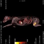
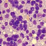
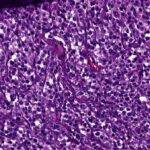
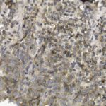
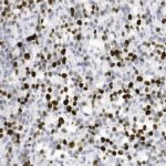
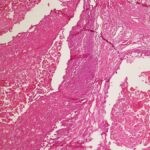
During the operative intervention, a mass in the lumbal area, macroscopically with muscle invasion, 5 cm in diameter, soft, with a lobulated cut surface, beige in color was discovered. The mass extracted from the hind leg distal was 2,5 cm in diameter, circumscribed, solid and localized on the skin. In the FDG PET/CT scan, on the left lateral side of 12. costa, upper and lower lumbal area, in left paramedian region there were dense hypermetabolic soft tissue nodules the most palpable of which was 23×17 mm in size and on the left femur distal area, a very dense hypermetabolic lesion (Image 1), an expansile soft tissue mass (the largest axial diameter was 80×39 mm). The examination of samples taken with fine needle aspiration showed neoplastic cell groups (Image 2) with narrow, pale basophilic cytoplasm, round, oval and some with notched nuclei, with distinct nucleoli, some with granular chromatin or mitotic figures. Histopathologically diagnosed as “non-epitheliotropic cutaneous lymphoma,” the sections of the lumbal mass on the skin consisted of groups of cells separated by broad or fine fibrovascular septa and covered by fibrous capsule inside the fat tissue, medium sized cells constituting cords, with a distinct cytoplasm, round or notched nuclei, granular chromatine, immunoblastic with small or distinct eosinophilic nucleolus or some lympholastic and lymphocytic with multiple nucleoli atypical lymphoid cells (Image 3). IHC staining showed positive for CD79a (Image 4) and negative for CD3 and that neoplastic cells were of B lymphocyte in origin. Ki-67 proliferation index was between 30%-50% (Image 5). Mass extracted from the skin of the hind leg showed pilomatrixoma characterized by multiple cystic hair follicules filled with keratin and ghost cells (Image 6).
Photographs (Left to right):
Image 1. Left femur distal hypermetabolic lesion PET/CT imaging
Image 2. Lumbo-dorsal subcutaneous mass, cytological smear, round, neoplastic cells, MGG staining, 400x
Image 3. Lumbo-dorsal subcutaneous mass, nonepitheliotropic cutaneous lymphoma, H&E staining, 200x
Image 4. Lumbo-dorsal subcutaneous mass, CD79a antibody positive, IHC staining, 200x
Image 5. Lumbo-dorsal subcutaneous mass, Ki-67 antibody positive, IHC staining, 200x
Image 6. Mass from the hind leg, pilomatrixoma, cystic follicules filled with keratin, H&E staining, 200x
Discussion and Results
Disease was observed to go into complete remission in 2 months after PET/CT scan findings showed femoral retention of B cell lymphoma and the patient’s treatment was modified when as a result of radiation and chemotherapy of Wisconsin protocol, the patient developed pancytopenia and was treated with chlorambucil-prednisolone and doxycycline. There were no reactions to radiotherapy. Femur functions remained intact. As of the time of this presentation (13 months after diagnosis) patient has survived without relapse. Pilomatrixoma, a benign epithelial tumor on the hind leg was discovered as the second neoplasia by coincidence and there was no relapse. In conclusion, cytology was determined to be an important and fast diagnostic tool in diagnosing neoplastic masses in dogs and IHC staining a significant tool in in determining the origins of the lymphoma cell as well as prognosis and treatment approach. In this case, IHC staining was used to show that the nonepitheliotropic cutaneous lymphoma had B cell origin. Although literature shows that nonepitheliotropic cutaneous lyphoma generally is solitary and localized on the skin, in our case, PET/CT showed multifocal retention. That is the reason why the case was considered to be worthy of discussion. In addition, we have shown that B cell lymphoma in dogs is sensitive to radiotherapy and chemotherapy treatment options and is a manageable disease.


